
Cytology
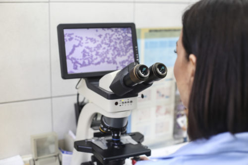
Cytology is the microscopic examination of cell samples. These samples can be collected from any area of the body. Cytology is often used to diagnose growths or masses (tumors) found on the surface of the body, but can also be used to assess bodily fluids, internal organs (e.g., liver, lung, lymph nodes, kidney), and abnormal fluids that may accumulate, especially in the chest and abdomen.
There are several ways to collect the cells depending on where the problem is and what type of tissue is involved.
Most commonly, fine needle aspiration or fine needle biopsy is performed to collect cells. This technique uses a sterile, fine gauge needle (a needle with a very small diameter) attached to an empty syringe. The needle is inserted into the middle of the tissue or pocket of fluid and the plunger of the syringe is pulled back to create suction and withdraw or aspirate cells from solid tissue, such as a skin lump, or to collect fluid from a site, such as a joint.
Biopsy
A biopsy is the surgical removal of a representative sample of tissue from a suspicious lesion. The biopsy is then processed and is examined under a microscope (see histopathology below). The most common biopsies are:
- punch biopsy – a small, circular piece of tissue is removed using a biopsy punch,
- wedge biopsy – a small slice or chunk of tissue is removed from the tumor or mass, or
- excision biopsy – the entire mass is excised or removed.
In some cases, a biopsy is taken knowing that surgical removal of the whole tumor is not possible. In other cases, a small biopsy can be useful to plan the surgical approach or determine if other treatments may be used, thereby increasing the chances of a successful outcome. However, in many cases, particularly when it is easy to get good surgical margins (area of ‘healthy’ tissue) around the mass, it is more appropriate to remove the whole mass. Your veterinarian will give your pet a local or general anesthetic in order to take samples.
Histopathology
Histopathology is the examination of samples of whole tissues and is performed on a solid piece of tissue that has been collected surgically.
The piece of tissue is prepared through a process called histology by preserving, thinly slicing or sectioning, and staining the tissue sample with dyes. Once prepared, the tissue sections are examined under the microscope by a veterinary pathologist. Histopathology focuses on the architecture of the tissue and provides more information about the tissue than cytology.
With this type of laboratory examination, the accuracy of a diagnosis is usually high. The veterinary pathologist can often offer an opinion on the likely course of the disease, what is called the prognosis. If the entire mass of tissue (the whole tumor) is submitted for examination, the pathologist may also offer an opinion as to whether the tumor has been completely removed i.e., if there are wide-margins (the amount of ‘healthy’ tissue surrounding the tumor). This information helps your veterinarian to decide the best course of treatment for your pet.